

الوظائف ذات الصلة
مستقبل G المرتبط بالبروتين: من إشارات الخلايا إلى اكتشاف الدواء
2026-07-29مستقبلات مرتبطة ببروتين G، أو GPCRs اختصارا، هي بروتينات غشائية تساعد الخلايا على الاستجابة للإشارات القادمة من خارج الخلايا. قد تشمل هذه الإشارات الهرمونات، والناقلات العصبية، والدهون، والببتيدات، بالإضافة إلى الضوء.
بينما هناك قواسم مشتركة واضحة في البنية السبعة عبر الأغشية في GPCRs، إلا أنها لا تعمل بنفس الطريقة. قد تعتمد نتائج التجربة على نوع الليغاند للمستقبل، وبيئة الخلية، ومستوى تعبير المستقبل، وشركاء الإشارة.
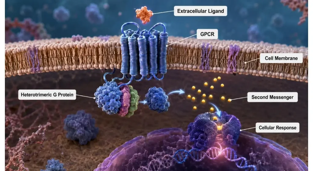
لذا، يجب على الباحثين تجاوز هذا القياس ل "تفعيل" المركب لمستقبل لفهم بيولوجيا GPCR. جميع المسارات، التوقيت، تنسيق الاختبار، التحكم ونموذج الخلية مهمة.
مستقبل بروتين G المقترن: ما هو؟
عدد المرات التي يعبر فيها GPCR نموذجي غشاء الخلية هو 7. ترتبط الأجزاء خارج الخلية منه بجزيئات الإشارة، والأجزاء داخل الخلية ترتبط ببروتينات G، والأريستينات، والكينازات وغيرها من جزيئات الإشارة.
تصنف GPCRs إلى عدد من الفئات الرئيسية حسب دليل IUPHAR/BPS لعلم الأدوية، مثل الفئة A، الفئة B، الفئة C، المستقبلات المجعدة، والالتصاق. الفئة A هي الأكبر، وتشمل أهدافا معروفة مثل الأدرينالين، الدوبامين، الأفيونات، والمستقبلات المسكارينية.
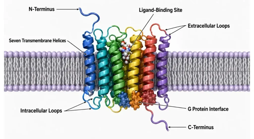
على الرغم من تشابه البنية، إلا أن GPCRs قادرة على التعرف على مجموعة متنوعة جدا من الإشارات. وهذا يمكنهم من الانخراط في الإدراك الحسي، وتنظيم القلب والأوعية الدموية، والتمثيل الغذائي، ووظائف المناعة، والمزاج، والحركة، وأكثر من ذلك بكثير.
كيف يتم تفعيل GPCRs؟
يرتبط الليجند بالمستقبل، مما يسبب تغير شكل المستقبل. نتيجة لهذا التغير التكوني، يستطيع المستقبل التفاعل مع بروتين إشارات داخل الخلية.
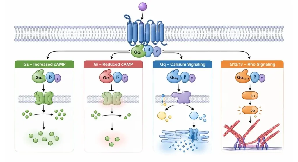
العديد من GPCRs تحتوي على بروتين G غير متجانس كشريك أول يتكون من وحدات ألفا وبيتا وغاما. يؤدي تنشيط المستقبلات إلى تبادل الناتج المحلي الإجمالي مع GTP في الوحدة ألفا. ثم تؤثر المكونات المنشطة على الإنزيمات اللاحقة، وقنوات الأيونات، والرسل الثانيين.
أكثر عائلات بروتينات G شعبية هي:
- Gs، الذي عادة ما يسبب زيادة في التيار الكهربائي الدوري
- الاسم الشائع المستخدم لإنزيم تقليل AMP الدوري هو gi/o.
- مسار Gq/11 الذي يشير إلى مسارات الفوسفوليباز C والكالسيوم
- يمكن التحكم في بروتين عائلة رو واستجابات الهيكل الخلوي بواسطة G12/13.
هذا الوصف المدرسي مفيد، لكن الخط هو شكل نادر من أشكال الإشارة! يمكن لمستقبل واحد أن يرسل إشارات عبر عدة بروتينات G، ويمكن أن يختلف نقل الإشارة الأمثل حسب الليجاند ونوع الخلية. يذكر GPCRdb أن المستقبلات يمكن أن تتفاعل مع بروتينات G، وكيناز GPCR، والأسريتينات، مما يؤدي إلى تكوين مجموعة متنوعة من مسارات الإشارة داخل الخلية.
أدوار الاعتقالات
بمجرد تنشيط بروتين G، لا تتوقف الإشارات من GPCRs.
كيناز GPCR قادر على فسفرة المستقبلات المنشطة. يمكن للأريستينات بعد ذلك التفاعل مع المستقبل، ومنع المزيد من الاقتران بين المستقبل وبروتينات G، وتسهيل استيطان المستقبلات، والمشاركة في مسارات أخرى لنقل الإشارة.
لذلك، لا يمكن اعتبار تجنيد ال arrestin ك "مفتاح إيقاف". اعتمادا على المستقبل والتجربة، يمكن أن يؤثر على التحريك أو إزالة التحسس أو إعادة تدوير أو تدهور المستقبل أو تعديل تأثيرات أخرى لاحقة.
نظرا لأن إشارات بروتين G واستدعاء التوقف هما مقاييس مهمة لعلم الأدوية الكامل للمركب، فغالبا ما يتم قياسهما معا.
تفحص المقالات في هذا القسم الطرق التي يمكن أن يؤدي بها نفس المستقبل إلى استجابات مختلفة
أجهزة GPCR ليست مجرد مفاتيح تشغيل/إيقاف.
يمكن العثور على روابط مختلفة ترتبط بمراحل تكوين مختلفة للمستقبل. يمكن أن يسبب نوع من الليغاند مستويات عالية من التنشيط في أحد مسارات بروتين G، بينما المسار الآخر قد يسبب زيادة تجنيد التوقف أو ملف الإشارات المختلف. يشار إليه عادة بالانتقائية الوظيفية/الإشارات المتحيزة.
خلفيات الخلايا مهمة أيضا. يمكن العثور على نفس المستقبل في مختبرين مختلفين، لكن يحتوي على كميات مختلفة من بروتينات G، أو الأسترينات، أو الكينازات، أو الإنزيمات، أو المستقبلات الداخلية في الخلايا.
مشكلة أخرى قد تكون مستوى تعبير المستقبلات. يمكن أن يعزز التعبير المفرط الاستجابات لمستويات النشاط المنخفضة و/أو يغير تفضيلات الاقتران، كما قد يؤدي إلى نشاط المحفز الجزئي في الخلية مقارنة بالأنسجة الأصلية.
لذا يجب اعتبار النتيجة مؤشرا على سلوك نظام تجريبي كامل وليس فقط على المستقبل.
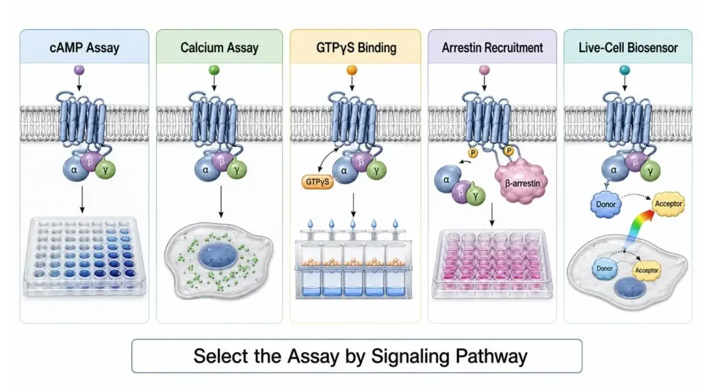
اختبار اختاري بناء على سؤال بيولوجي
لا يمكن لاختبار GPCR واحد أن يجيب على جميع الأسئلة.
| طريقة الفحص | القراءة الرئيسية | الاستخدام الشائع | الاعتبار الرئيسي |
| اختبار cAMP | التغير في cAMP الخلوي | مستقبلات Gs وGi مرتبطة | cAMP القاعدي وطريقة التحفيز |
| اختبار الكالسيوم | تغير الكالسيوم داخل الخلية | المستقبلات المرتبطة ب Gq | إشارة سريعة وتوقيت دقيق |
| ربط GTPγS | تنشيط بروتين G | تقييم المنشطات المباشرة | جودة تحضير الأغشية |
| الاعتقال في التجنيد | تفاعل المستقبلات مع التوقف | إزالة التحسس وتحيز المسار | التفسير باستخدام اختبار بروتين G |
| المستشعر الحيوي للخلايا الحية | حركيات الإشارات في الوقت الحقيقي | التنشيط وديناميكيات المحولات | تعبير المستشعر وتصميم الاختبار |
اختبارات cAMP
عادة ما يتم قياس المستقبلات التي تحتوي على بروتينات Gs أو Gi باستخدام cAMP. قد تؤدي استجابة Gs إلى زيادة cAMP، ويمكن أن تشير استجابة G. إلى تثبيط إنتاج cAMP المحفز.
تستخدم هذه الاختبارات عادة في دراسات التركيز والاستجابة الذهنية، وفي دراسات توصيف المنشطات، وفي دراسات المضادة. في تجارب المضاد، عادة ما يتم تطبيق تركيز من المنشط بعد تعرض المركب الاختبار للخلايا.
اختبارات الكالسيوم
يمكن دراسة المستقبلات المرتبطة ب Gq من خلال اختبارات داخل الخلية للكالسيوم. يمكنها أن تعطي قراءة سريعة وعالية الإنتاجية، لكن الاستجابة قد تكون قصيرة المدة، وتعتمد بشكل كبير على كيفية التعامل مع الخلايا، وتحميل الأصباغ، وتعبير المستقبلات، ووقت القياس.
ربط GTPγS
اختبارات GTPγS هي من أوائل الأحداث التي يتم قياسها في عملية تنشيط بروتين G وغالبا ما تستخدم لتأكيد نشاط المنشط في تحضيرات الغشاء. نظرا لأن الاختبار قريب جدا من تفاعل المستقبل مع بروتين G، يمكن أن يقلل من بعض التضخيم الذي يلاحظه في القراءات اللاحقة.
توظيف الاعتقال
يمكن استخدام اختبارات أريستين للتحقيق في تحيز المسارات، الإشارة الخاصة بالليغاند، تنقل المستقبلات، وإزالة التحسس للمستقبلات. من الأفضل استخدامها مع اختبار بروتين G وليس كمؤشر عام على تنشيط المستقبل.
حساسات الخلايا الحيوية الحية
تقنيات BRET وFRET (وغيرها من طرق الاستشعار الحيوي) يمكنها تتبع أحداث الإشارات في الخلايا الحية كدالة للزمن. أكبر نقطة بيع في هذا هو المعلومات الحركية: يستطيع الباحثون قياس متى تبدأ الإشارات، ومتى تبلغ ذروتها ثم تنخفض، لأنهم يقيسون أكثر من نقطة نهاية.
قم بإجراء تجربة دقيقة ل GPCR
الاختبار الجيد يعتمد على البيولوجي وليس على الجهاز.
أولا، تأكد من أن المستقبل معبر عنه وموقعه في الموقع الصحيح في غشاء الخلية. قد لا يكون انخفاض الاستجابة نتيجة لانخفاض نشاط الليغاند، بل نتيجة ضعف التعبير أو الحركة في المستقبلات.
ثانيا، أضف أجهزة تحكم مناسبة. عادة ما تكون هناك حاجة إلى منبه معروف، ومضاد معروف، وأدوات تحكم للمركبات، وخلايا غير منقلة، وتحكم مستقل عن المستقبل للفحص اللاحق.
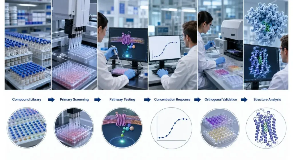
وأخيرا، قم بإجراء منحنى تركيز واستجابة كامل. تقييم تركيز واحد فقط من المركب لن يكون مقياسا موثوقا للفعالية أو التنهيد الجزئي أو السمية أو التداخل غير النوعي مع الاختبار.
هناك حاجة أيضا إلى توحيد التوقيت. بمجرد أن تحصل على إشارة الكالسيوم، خلال ثوان قليلة، ثم بعد ساعات قليلة تقيس التعبير الجني، لا يمكنك ببساطة مقارنة الاثنين. يمكن استخدام اختبارات تقيس نفس المستقبل لقياس مراحل مختلفة من الاستجابة.
وأخيرا، تحقق من النتائج المهمة باستخدام النهج المتعامد. إذا وجد أن المركب نشط في اختبار cAMP، من الأفضل تأكيده باختبار مفيد آخر، أو اختبار الربط أو باختبار إيجابي خاص بالمستقبل. يشير دليل إرشادات الفحص إلى أن التحقق الدقيق، ومراقبة الجودة، والتوصيف الدوائي لنتائج GPCR في الانتقال نحو تحسين الرصاص سيكون موصى به.
شهادات GPCR لاكتشاف الأدوية
تستمر GPCRs في لعب دور رئيسي في اكتشاف الأدوية لأنها تشارك في العديد من جوانب الفسيولوجيا وتعبر عنها على السطح على الخلية. يقدر GPCRdb أن 25٪ من الهرمونات البشرية تشارك استجابة لأعضاء هذه العائلة المستقبلية، وأن حوالي ثلث جميع الأدوية تستهدف أعضاء هذه العائلة.
بالنسبة للبرامج التقليدية، كانت أهداف البحث الرئيسية إما منبهات أو مضادات ترتبط بموقع ربط الرابطة الرئيسي. كما تجري حاليا دراسة في المعدلات الفضائية، والروابط المنحازة للمسار، وانتقائية المستقبلات والمحولات، وحركية الارتباط، وتصميم المركبات القائمة على البنية.
مع ظهور علم الأحياء البنيوي، أصبح هذا أكثر قابلية للتطبيق. يتضمن GPCRdb الآن أدوات لمقارنة مواقع الارتباط وحالات تنشيط GPCRs، بما في ذلك هياكل المستقبلات التي تم تحديدها تجريبيا، ومركبات المستقبلات–الليجاند، ومركبات بروتينات المستقبلات–G.
يجب أن يخضع المركب ذو الهياكل الجيدة لاختبار وظيفي حتى مع وجود هياكل جيدة. يمكن للجزيء أن يرتبط بهدف مناسب ذو ألفة ارتباط قوية دون أن يثير الاستجابة الخلوية المقصودة، أو بدلا من ذلك، قد يتفاعل الجزيء مع المسار المطلوب ولكن ليس بالطريقة الصحيحة.
استنتاج
من الأفضل اعتبار مستقبلات بروتين G ليست مجرد مفتاح غشاء، بل نظام إشارات مرن.
يتغير شكل المستقبل استجابة لارتباط الرابط، ويجد المستقبل شركائه في الخلية، والبيئة تحدد مسار الإشارة. لهذا السبب من المهم النظر في اختيار الاختبارات، وتعبير المستقبلات، وتوقيت المسار، والضوابط، والتحقق المتعامد.
غالبا ما يتم التوصل إلى أفضل الاستنتاجات لأبحاث GPCR واكتشاف الأدوية نتيجة لمزيج من المعلومات الهيكلية وأكثر من اختبار وظيفي واحد.
الأسئلة الشائعة
س1. ما هي الوظيفة الرئيسية ل GPCR؟
GPCR هو مستقبل سطح الخلية يستجيب لإشارة خارج الخلية وينتج استجابة داخل الخلية. قد يكون الجواب عبر بروتينات G، أو الريتينات، أو الرسل الثاني، أو قنوات الأيونات أو تنظيم الجينات.
س2. هل جميع GPCRs يتم تنشيطها بواسطة نفس بروتين G؟
لا. يمكن لمستقبلات مختلفة أن تفضل تنشيط بروتينات G مختلفة (Gs، Gi/o، Gq/11، G12/13 أو شركاء إشارات متعددين).
س3. ما هو GPCR اليتيمي؟
GPCR اليتيم هو مستقبل لا يكون من المؤكد له الليغاند المنشط الطبيعي. لا تزال هذه المستقبلات ذات أهمية كبيرة للأبحاث الأساسية وتطوير الأدوية.
س4. فأي اختبار يجب استخدامه لفحص GPCR؟
سيعتمد "أفضل" الاختبار على السؤال الذي يتم معالجته ومسار الإشارة للمستقبل. يمكن قياس جوانب مختلفة من الاستجابة بواسطة اختبارات cAMP، والكالسيوم، وGTPγS، وأريستين، واختبارات المستشعرات الحيوية للخلايا الحية.
س5. ما السبب الذي يدفع الباحثين لاستخدام أكثر من اختبار واحد؟
يمكن لاختبار واحد قياس مسار واحد فقط أو وجود تداخلات خاصة بالتنسيق. إذا أمكن إثبات من خلال اختبار متعامدة أن التأثير المرصود مرتبط بالفعل بالمستقبلات، فمن الممكن الاستنتاج أن هذا هو تأثير حقيقي للمستقبل.
