
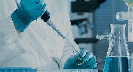
خدمة المجهر الإلكتروني البارد
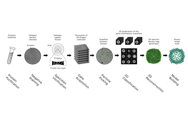
المجهر الإلكتروني البارد (cryo-EM) هو تقنية متقدمة في علم الأحياء البنيوي تتيح للعلماء تصور التفاصيل الدقيقة للجزيئات الكبيرة البيولوجية. يعتمد التجميد الكهرومغناطيسي على المجهر الإلكتروني الناقل بدقة قريبة من الذرة. زيادة الطلب في السوق في العديد من التطبيقات الطبية الحيوية، مثل تطوير الأدوية، والأجسام المضادة، واللقاحات، وما إلى ذلك.
مزايا التجميد الكهرومغناطيسي مقارنة بعلم البلورات بالأشعة السينية
على الرغم من أن حيود الأشعة السينية لا يزال يمكن أن يصل إلى دقة أعلى قليلا، إلا أن التجميد الكهرومغناطيسي يظهر مزايا كبيرة:
- كمية قليلة من العينة مطلوبة.
- لا حاجة للتبلور.
- لا حاجة لمصدر الأشعة السينكروترونية السينكروترونية.
- الحفاظ على الحالة شبه الأصلية.
- تقييم المرونة.
- مناسبة للجسيمات التي يصعب تبلور مثل البروتينات الغشائية أو التجمعات فوق الجزيئية (مثل الفيروس).
- يمكن حل تباين التكوينات في عينة واحدة.
- مستوى منخفض من النقاء مقبول.
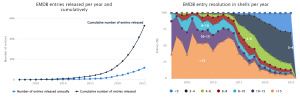
النمو الأسي في عدد المشاركات في EMDB:
الحلول الهيكلية
- تقييم ملاءمة العينة – الصبغة السلبية
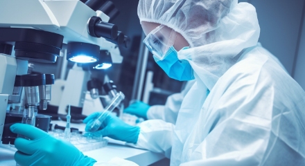
قبل تخصيص موارد مهمة من المجهر في التجميد الكهرومغناطيسي، يتم عادة إجراء فحص الصبغة السالبة في درجة حرارة الغرفة والجهد المنخفض. تسمح هذه الخطوة بتقييم ملاءمة العينة، بما في ذلك التجانس، والتجميع، وشكل الجسيمات، وحجمها، والتوزيع، باستخدام عامل تلوين عالي الكثافة الإلكترونية.
يعد التلوين السلبي خطوة حاسمة لمراقبة الجودة لتقييم ما إذا كانت العينة مناسبة لجمع بيانات التجميد والEM لاحقا.
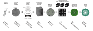
- تحديد البنية عالية الدقة – تحليل الجسيمات المفردة (SPA)
عادة ما يولد جمع بيانات التجميد الكهرومغناطيسي حوالي 10⁴ إلى 10⁶ إسقاطات ثنائية الأبعاد فردية للجسيمات الملتقطة في اتجاهات مختلفة. يتم محاذاة هذه الإسقاطات حسابيا وتصنيفها من خلال سير عمل معالجة الصور لإعادة بناء خريطة كثافة ثلاثية الأبعاد بدقة دون النانومترية أو قريبة من الذرة.
يتم بناء نموذج ذري وتطويره والتحقق منه لاحقا ضمن خريطة الكثافة المعاد بناؤها، مما يوفر رؤى هيكلية حول الآليات الوظيفية للجزيء الكبير البيولوجي المعني.
- التحليل البنيوي في الموقع – التصوير المقطعي بالإلكترون البارد (Cryo-ET)
يتيح التصوير المقطعي بالإلكترون البارد التصور ثلاثي الأبعاد للهياكل البيولوجية في سياقها الخلوي أو الفيروسي الأصلي. هذا النهج مناسب بشكل خاص لدراسة التجمعات الجزيئية الكبيرة الكبيرة، والمجمعات المرتبطة بالغشاء، وآليات العدوى الفيروسية، حيث يقدم معلومات مكانية وتنظيمية لا يمكن الوصول إليها بطرق الجسيمات الفردية.
سير العمل الخدمي
استشارة المشروع → توقيع اتفاقية عدم الإفشاء → تأكيد → العينات التي تلقت → فحص الجودة → فحص البقع السلبية → جمع بيانات التجميد-EM → معالجة البيانات → تسليم التقارير.
البيانات والنتائج المخرجات
يتم توفير توفر البيانات الكاملة طوال دورة حياة المشروع. تشمل المواد المقدمة أفلام التجميد الخام مع ملفات مرجعية للكسب، وملفات بيانات وسيطة في مراحل مختلفة من معالجة البيانات، وخرائط الكثافة ثلاثية الأبعاد النهائية مع مقاييس الدقة والجودة، ونماذج الإحداثيات الذرية عند الاقتضاء، وتقارير تحقق متقاطع مثل تقييمات MolProbity.
يمكن تسليم جميع البيانات عبر أجهزة تخزين محمولة أو منصات سحابية آمنة.
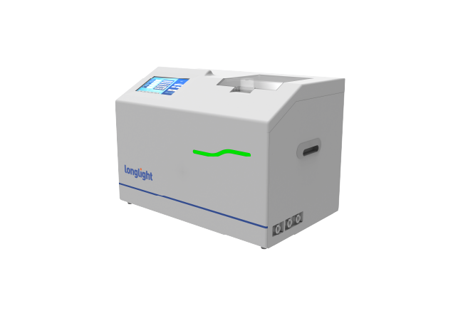
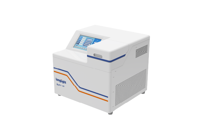
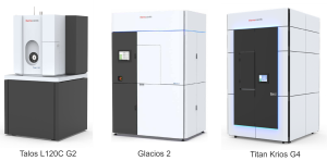
ثلاثة معدات رئيسية
تدعم خدماتنا للبريد-EM من خلال الوصول إلى منصات بحث علمي متقدمة مجهزة بأحدث الأجهزة.
تشمل الأنظمة المتاحة تالوس L120C G2، منصة مجهر إلكتروني نقل للمبتدئين وفحص الكهرومغناطيسية بالبرود الكهرومغناطيسي؛ الأنهار الجليدية 2، نظام تجميد كهربائي يعمل بجهد 200 كيلو فولت محسن لسير عمل الجسيمات المنفردة والتصوير المقطعي الروتيني؛ و تايتان كريوس G4، منصة رائدة بقدرة 300 ك.ف كريو-EM مصممة لتحقيق أقصى استقرار وأتمتة وسرعة نقل وإمكانية دقة.
تدعم هذه المنصات مجتمعة فحص الشبكة التبريدية، وتحليل الجسيمات الفردية، والتصوير المقطعي بالإلكترونات بالتبريد، وتطوير الطرق المتقدمة عبر مجموعة واسعة من تطبيقات علم الأحياء الهيكلي.
لماذا لونغلايت؟
نقدم خبرة قيمة وناضجة في المجهر الإلكتروني البارد من خلال الوصول إلى منصات بحث علمي متقدمة والتعاون مع خبراء الصناعة والمستشارين العلميين الأكاديميين. تركز خدماتنا على الشفافية الكاملة وشمولية تسليم البيانات، وتشمل البيانات الخام، وملفات المعالجة الوسيطة، وخرائط الكثافة النهائية، والنماذج الذرية، وتقارير التحقق.
من خلال دمج الأجهزة المتقدمة، وسير عمل معالجة البيانات ذات الخبرة، وجداول زمنية متوازنة في التسليم، نوفر حلول هيكلية عالية الجودة وموثوقة لتقنية التجميد المغناطيسي المغناطيسي الدقيقة مصممة خصيصا لتلبية احتياجات البحث المتنوعة.
الأسئلة الشائعة.
س1: كم من العينة يحتاج العملاء إلى تحضيرها؟
- الصبغة السلبية:
تركيز أدنى ~1 جم/لتر
الحد الأدنى للحجم ~100 ميكرولتر.
- SPA مع بروتينات قابلة للذوبان:
تركيز أدنى ~1 جم/لتر
حجم أدنى ~100 ميكرولتر.
- SPA مع بروتينات الغشاء:
تركيز أدنى ~1 جم/لتر
حجم أدنى ~100 ميكرولتر.
ليتم مناقشتها وتعديلها إذا لزم الأمر
س2: ما هي متطلبات مخزن العينة؟
- نطاق الحموضة: 6.0-8.5
- تركيز الملح <200 ملليمولار
- انخفاض الجلسرول (كم السعر؟ رقم التأثير: 10.1107/S2053230X24002553)
- أزيد منخفض (كم الكمية؟ المرجع الداعم مفقود)
س3: ما هي عملية الخدمة النموذجية؟
استشارة المشروع → توقيع اتفاقية عدم الإفراج → تأكيد → عينة → فحص الجودة → → السلبية جمع البيانات → معالجة البيانات → تسليم التقارير.
س4: ما هو متوسط وقت التسليم؟
- التصبغات السلبية: من أسبوع إلى أسبوعين.
- سبا، النتائج الأولية: 6-8 أسابيع.
- SPA، نموذج عالي الدقة: 2-3 أشهر.
تشيب-seq
يدرس ChIP التفاعل بين الحمض النووي والبروتينات ، ويتم دمج ChIP-seq مع تسلسل الجيل التالي للكشف عن مواقع الحمض النووي في الجينوم التي ترتبط بعوامل نسخ / هيستونات محددة ، والتي تستخدم لدراسة التفاعل بين البروتينات والكروماتين.
علم الجينوم
ركزت Longlight على التشخيص الجزيئي والبيولوجيا الجزيئية ، وقد أطلقنا بعض الأدوات والمواد الاستهلاكية ذات الصلة ب NGS ، وخاصة الموجات فوق الصوتية المركزة ، ملتزمة بتعزيز تطوير علم الجينوم ، لخدمة القضية الطبية البشرية بشكل أفضل.
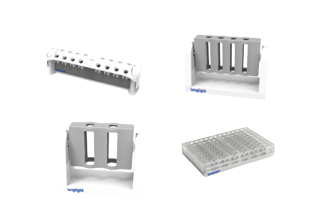
المواد الاستهلاكية والأطقم
يوفر Longlight أنواعا مختلفة من هلام الاغاروز مسبقة الصب ، وزبال الحمض النووي ، وأنبوب Qubit ، ومجموعات استخراج الحمض النووي ، ومجموعات تحضير المكتبة. وفي الوقت نفسه، يمكن استخدام كل هذه المنتجات على نطاق واسع في البحث العلمي الأساسي والمستحضرات الصيدلانية الحيوية وسيناريوهات التطبيق الأخرى.
تواصل معنا


دعم الخط الساخن
للحصول على استجابة أسرع، يمكنك التواصل معنا عبر الهاتف: 86 0755-86727654