
الوظائف ذات الصلة
نظام تصوير الأحماض النووية: كيف تحل المختبرات النطاقات الضعيفة وفجوات التتبع
2026-03-16أصبح اختيار نظام تصوير الأحماض النووية قرارا عمليا للمختبرات التي سئمت من مشكلتين متكررتين: التقاط النطاق الضعيف الذي لا يدعم تفسيرا واثقا، وسجلات الصور المجزأة التي تجعل المراجعة الداخلية أو سير العمل المنظم أصعب مما ينبغي.
(تقنيات تحليلية لاكتشاف الأحماض النووية والبروتينات مع حساسية جزيء واحد)
A وحدة توثيق GEL لم يعد مجرد صندوق لالتقاط الصور بعد الرحلان الكهربائي. الآن يقع عند تقاطع حساسية الصور، وسلامة المستخدم، وسرعة سير العمل، وتتبع البيانات. في سير عمل الأحماض النووية والبروتينات، تكون الأهداف منخفضة الوفرة والإشارات الضعيفة شائعة بما يكفي لتؤثر حساسية التصوير بشكل مباشر على ما إذا كانت النتيجة قابلة للاستخدام أو ما إذا كان يجب تكرار التشغيل. على سبيل المثال، يبرز Bio-Rad اكتشاف الأهداف منخفض الوفرة كتحد جوهري في التصوير في سير عمل البلوت والجل.
لماذا tالسوق يتجاوز توثيق الجل الأساسي
لا تزال العديد من المختبرات تتعامل مع قيود أنظمة التصوير القديمة. الأعراض المعتادة مألوفة:
• تختفي النطاقات الضعيفة في ضوضاء الخلفية
• وضع العلامات اليدوية على السلم يبطئ التقارير
• المستخدمون المختلفون يحتفظون بالملفات بطرق غير متسقة
• تدفقات العمل التي تعمل فقط بالأشعة فوق البنفسجية تضيف مخاوف تلف الحمض النووي أثناء استئصال الجل
• يتم التقاط الصور، ولكن لا تدار بطريقة تدعم مراجعة التدقيق
وهذا مهم لأن التجربة في البداية غالبا ما تكون جزءا فقط من التكلفة. عندما يكون التصوير غير متسق، يكون الخسارة الحقيقية هي وقت الفني، والتحليل المتأخر، وانخفاض الثقة في تفسير العينات.
أصبح تصوير الضوء الأزرق أكثر أهمية في هذا السياق. يشير ثيرمو فيشر إلى أن الضوء الأزرق يسبب ضررا طفيفا في الحمض النووي مقارنة بالأشعة فوق البنفسجية ويمكن أن يحسن كفاءة الاستنساخ عند الحاجة إلى استعادة الحمض النووي. كما يشير إلى أن التعرض للأشعة فوق البنفسجية يمكن أن يقلل من كفاءة الاستنساخ، وهو أمر عملي في سير العمل الذي يتضمن الاسترداد والاستنساخ في مرحلة لاحقة.
إجابة أكثر عملية to الفرق الضعيفة aتأخيرات سير العمل
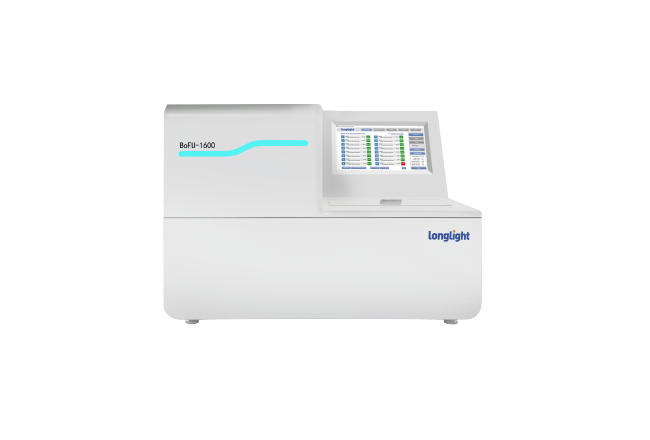
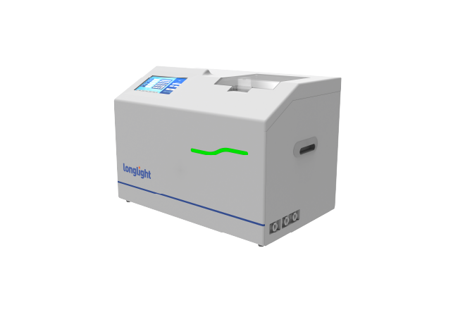
تم بناء تقنية لونغلايت GI-200 حول احتياجات المختبرات التي ترغب في منصة واحدة تغطي كل من التصوير الروتيني وتصور العينات الأكثر تطلبا.
تشمل المواصفات الرئيسية:
• كاميرا CMOS بالأبيض والأسود عالية الحساسية بدقة 6.3 ميجابكسل
• نسبة الإشارة إلى الضوضاء 66 ديسيبل
• مساحة تصوير 175 مم × 230 مم
• إثارة عابرة للأبيض، عبر الأشعة فوق البنفسجية، وعبر الأزرق
• شاشة لمس مدمجة بحجم 12.1 بوصة
• معالجة الصور على متن السفينة والتوثيق الرقمي
• دعم قطع هلام خارجي مع لوحة قطع واقية من الأشعة فوق البنفسجية
من منظور الشراء، النقطة المهمة ليست رقم الميجابكسل وحده. إنه مزيج من الحساسية، وانخفاض الضوضاء الخلفية، والأداء البصري المسيطر عليه. التصوير عالي الحساسية مهم عندما تحتاج المختبرات إلى التقاط أحزمة حمض نووي أو بروتينات ضعيفة دون تعريض إشارات أقوى بشكل مفرط. وهذا مهم بشكل خاص في المختبرات الأساسية المشتركة، ومختبرات التدريس، ومختبرات الأحياء الجزيئية، وفرق البحث التطبيقي التي تعمل عبر أشكال مختلفة من الألوان والجل.
(تقنيات تحليلية لاكتشاف الأحماض النووية والبروتينات مع حساسية جزيء واحد)
حيث يتناسب GI-200 بشكل أفضل مع الاستخدام الحقيقي في المختبر
يجب أن يقلل نظام التصوير القوي من خطوات التعامل، وليس أن يخلق المزيد منها. يفعل GI-200 ذلك بعدة طرق.
- تصوير مدمج بدلا من سير عمل مجزأ
يجمع التصميم الشامل بين التصوير والمعالجة والمشاهدة على شاشة لمس مقاس 12.1 بوصة. هذا يقلل الاعتماد على جهاز كمبيوتر خارجي لكل مهمة أساسية، مما يمكن أن يبسط وضع الطاولة والاستخدام اليومي. بالنسبة للمشترين الذين يجهزون مختبرات صغيرة أو غرف متعددة المستخدمين، غالبا ما يكون هذا النوع من سير العمل المستقل أكثر فائدة من إضافة محطة كمبيوتر أخرى.
- دعم أفضل لاكتشاف الإشارة الضعيفة
تستخدم كاميرا GI-200 من لونجلايت كاميرا CMOS عالية الحساسية بدقة 6.3 ميجابكسل مع أداء تصوير منخفض الضوضاء. عمليا، هذا هو نوع المواصفات التي يجب أن يبحث عنها المشترون عندما تقوم فرقهم بتشغيل عينات منخفضة الوفرة أو عندما تحتاج إلى فصل واضح بين الخطوط في ظروف الإضاءة المنخفضة. الحساسية ليست ميزة فاخرة في هذه الفئة. وهو أحد العوامل الرئيسية التي تحدد ما إذا كانت صورة الهلام جاهزة للنشر، أو جاهزة للمراجعة، أو مفيدة جزئيا فقط. هذا التركيز الأوسع في الصناعة على الحساسية يتماشى مع كيفية مناقشة الموردين الرئيسيين لتحديات التصوير في الإشارة المنخفضة.
- الضوء الأزرق، الأشعة فوق البنفسجية، والضوء الأبيض في نظام واحد
مرونة الصبغة هي مشكلة شراء أخرى تهم أكثر مما تشير إليه العديد من أوراق المنتج. يدعم GI-200 الضوء فوق البنفسجي العابر والأزرق والأبيض، مما يمنح المختبرات توافقا أوسع عبر تطبيقات الأحماض النووية والبروتينات. تشير ثيرمو فيشر إلى أن أصباغ مثل SYBR Safe يمكن مشاهدتها بضوء أزرق أو UV، بينما يفضل الإثارة بالضوء الأزرق عندما يكون تقليل تلف الحمض النووي أمرا مهما.
بالنسبة للموزعين ومديري المشتريات، تساعد هذه المرونة في تقليل الحاجة إلى وحدات منفصلة مخصصة لمصدر ضوء واحد فقط أو عائلة بقع واحدة.
لماذا التتبع الآن a متطلبات الشراء
نقطة ألم أخرى تم تجاهلها في تصوير الجل ليست التقاط الصورة، بل حوكمة الصورة.
يشمل GI-200:
• إدارة المستخدمين القائمة على الأدوار
• عرض السجلات المصنف
• سجل تدقيق كامل
• إدارة البيانات من أجل التتبع
• التعليق التلقائي على حجم نطاق السلم
وهذا مهم لأن العديد من المختبرات تحت ضغط لتحسين جودة التوثيق الداخلي، خاصة في البيئات التي يستخدم فيها عدة مشغلين نفس الجهاز. يمكن للنظام الذي يسجل من قام بماذا، ومتى تم التقاط الصورة، وكيفية التعامل مع البيانات أن يقلل من الالتباس أثناء المراجعة ويجعل سير العمل أكثر قابلية للدفاع.
وظيفة التعليق التلقائي على العلامة تعالج أيضا عبئا أصغر لكنه حقيقي جدا في سير العمل. وضع العلامات اليدوية على السلم متكرر، معرض للأخطاء، وغير متسق بين المستخدمين. يمكن أن يوفر التعرف التلقائي والتصنيف الوقت مع توحيد إعداد التقارير.
خيار أكثر أمانا fأو سير عمل إزالة الجل
السلامة هي سبب آخر يجعل المشترين يبتعدون عن أنظمة أحادية الوضع القديمة. يعتبر تصوير الضوء الأزرق ذا قيمة واسعة لأنه يقلل من خطر تلف الحمض النووي أثناء سير عمل التعافي، بينما يمكن لإعدادات المشاهدة الأكثر أمانا أن تقلل أيضا من مخاوف التعرض المباشر للأشعة فوق البنفسجية لدى المستخدمين. توصي شركة ثيرمو فيشر تحديدا بطرق الضوء الأزرق لتحسين حماية الحمض النووي وتلاحظ قيمتها أثناء أعمال الاستئصال.
يدعم GI-200 قطع الجل الخارجي ويشمل لوحة قطع واقية من الأشعة فوق البنفسجية قياسية، وهي ميزة عملية للمختبرات التي تقوم بإزالة الأشرطة بانتظام وترغب في حماية أفضل للمشغلين.
لماذا تستحق تقنية الإضاءة الطويلة a نظرة أقرب
بالنسبة للمشترين الذين يقارنون منصات توثيق الجيلام، يبرز GI-200 لأنه يعالج المشاكل التي تبطئ العمل المخبري فعليا:
• يحسن رؤية النطاقات الضعيفة
• يدعم مصادر ضوء متعددة وأنظمة صبغ
• يقلل من جهد التعليق اليدوي
• يعزز التتبع ومساءلة المستخدمين
• يساعد المختبرات على تحقيق التوازن بين الأداء والسلامة وكفاءة التكاليف
وهذا يجعله مناسبا للمختبرات التي تبحث عن نظام تصوير حمض نووي أكثر اكتمالا، وليس مجرد كاميرا هلام بسيطة.
في سوق لا تزال العديد من الأجهزة تجبر المختبرات على الاختيار بين الحساسية والمرونة والتحكم في التوثيق، يقدم GI-200 من Longlight Technology إجابة أكثر توازنا. بالنسبة لفرق البيولوجيا الجزيئية، والمختبرات الأكاديمية، ومرافق الاختبار، وشركاء القنوات الذين يخدمون هؤلاء المستخدمين، فإن هذا التوازن غالبا ما يحول نظام التصوير من شراء روتيني إلى ترقية سير العمل.