

الوظائف ذات الصلة
الرحلان الكهربائي الهلامي الأفقي للحمض النووي البلازميدي: فصل الأشكال الملفوفة والمحفوفة والخطية
2026-06-19الرحلان الكهربائي للحمض النووي هو طريقة شائعة الاستخدام في علم الأحياء الجزيئي. أحد التطبيقات التي يتطلب فيها أعلى مستوى من الفصل هو تحديد الأشكال الطوبولوجية الثلاثة الرئيسية للحمض النووي البلازميدي، وهي الأشكال الملفوفة الفائقة، والمثقوبة (الدائرية المفتوحة)، والأشكال الخطية. سنوضح كيف يوفر نظام HE-50 الجديد للهلام الأفقي عزل البلازميدات ومراقبة الجودة مما يسمح بفصل وتكرار أكبر.
[خمسة اعتبارات لعملية الرحلان الكهربائي للهلام بحمض الحمض النووي | ثيرمو فيشر ساينتيفك - الولايات المتحدة]
طوبولوجيا البلازميدات: ثلاثة أشكال
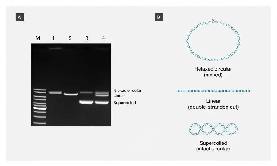
حمض البلازميد النووي غير منتظم وهو مزيج من ثلاثة أشكال طوبولوجية. في سياق العزل، ستنتج هذه الأشكال معدلات هجرة مختلفة أثناء الرحلان الكهربائي للحمض النووي.
• شكل الفائق الملفوف (SC): تعتبر البلازميدات الملفوفة الفائقة أسرع أنواع مهاجرة في هلام الأغاروز.
• الشكل المقشر (الدائري المفتوح/الهيكلي): الترتيب الدائري للحمض النووي البلازميدي الذي يحتوي على كسر لسلسلة واحدة أصبح الآن مسترخيا وبالتالي مشوشا. وبسبب الحجم الفعال الأكبر، فإن هذا الشكل يهاجر أبطأ من الحمض النووي الملفوف الفائق.
• الشكل الخطي: كسر كلا الخيطين التساهميين للبلازميد يؤدي إلى حمض نووي خطي. هذا النوع من الحمض النووي للبلازميدات له بنية على شكل قضيب، وهجرته أبطأ من السوبرلفائد، لكنها أسرع من الخدش.
عند تحليل جودة حمض البلازميدات النووي المستخدم في تصنيع لقاحات بلازميدات الحمض النووي، تشترط الجهات التنظيمية العليا أن يكون التحضير على الأقل 80٪ من البلازميد في الشكل الملفوف الفائق.
أهمية طوبولوجيا البلازميدات في البحث والأدوية الحيوية
في الحمض النووي للبلازميد، يؤثر شكل الأشكال المتشابكة على عدد من العوامل في العمليات ذات الصلة المتعلقة باللقاحات:
• التحويل إلى الخلايا الثديية: الشكل الفائق الملفوف من حمض البلازميد هو الأكثر فعالية في نقل الخلايا الثديية مقارنة بالأشكال المسترخية والخطية.
• إنتاج لقاح mRNA: الحاجة إلى قوالب بلازميد خطية للنسخ الصناعي (IVT) تؤدي إلى ملوثات دائرية مفتوحة للبلازميدات، مما يؤثر على الإنتاجية والقوام.
• تصنيع ناقلات العلاج الجيني: البلازميدات الملفوفة عالية الجودة ضرورية لإنتاج ناقلات التغذية الملحقة (AAV).
• مراقبة الاستقرار: تؤثر ظروف التخزين ومدة التخزين على استقرار البلازميدات مع زيادة تكرار الأشكال المكسورة أو الخطية.
التقدم البحثي الحديث في تحليل الحمض النووي للبلازميدات
تؤكد اثنتان من الدراسات الحديثة على أهمية الفصل عالي الدقة للحمض النووي للبلازميدات:
1. تسريح البلازميدات بوساطة زوري (2025)
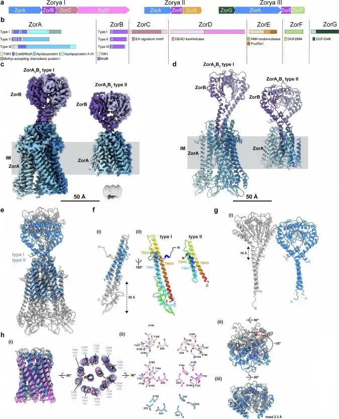
دراسة نشرت في مجلة Nature Communications (مارس 2025) استهدفت أنظمة الدفاع البكتيرية وأظهرت السيطرة على طوبولوجيات البلازميدات. وصفت مجموعة البحث تحت معهد زوريا تحولا بطيئا للحمض النووي البلازميدي pSG483 الملفوف بشكل فائق اللف إلى أشكاله المشققة والخطية بواسطة بروتين ZorE. لوحظ أن هذه العملية كانت تدريجية، وتم تحليل التحويل كميا باستخدام ImageJ لإجراء دراسة كثافة سكانية. لوحظ أن البلازميدات الملفوفة الفائقة تم خدشها تدريجيا بواسطة ZorE (768 نانومتر) بعد حضانة لمدة 60 دقيقة عند 37 درجة مئوية.
[اقتطاع البلازميدات بوساطة زوري (2025)]
*"تم إيقاف التفاعل بإضافة EDTA وSDS، وتم فصل المنتجات باستخدام رحلان كهربائي هلام في محلول 1× TAE، وجل أغاروز بنسبة 1.4٪، وتم صبغها بعد الطلاء ب eb."* — نيتشر كومينيكيشنز، 2025
وقد وجد أن هذا الاكتشاف كان حاسما بالنسبة لدراسات تفاعل شظايا الحمض النووي والبروتين باستخدام أنظمة الهلام.
2. الحمض النووي غير B وبروتينات إصلاح الحمض النووي (2025)
أظهر منشور PNAS من المعاهد الوطنية للصحة في يناير 2025 أن إدخال تكرارات الثنائي النيوكليوتيد في بلازميدات pUC18 له تأثير CIS على ارتباط بروتينات إصلاح عدم التطابق Mlh1-Pms1. اختبرت EMSA ارتباط البروتينات بتركيزات تتراوح بين 25 نانومول إلى 400 نانومول في كل من الحمض النووي الملفوف والخطي. الحمض النووي غير الموجود في الشكل B يعطل وظائف إصلاح البروتين ويضيف إلى فهم عدم استقرار الحمض النووي.
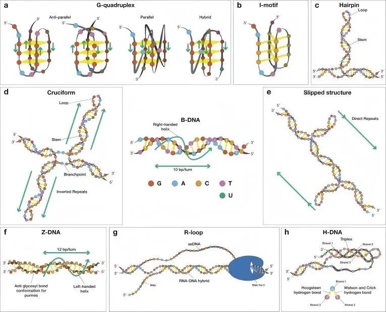
[الحمض النووي غير B وبروتينات إصلاح الحمض النووي (2025)]
اعتبارات مهمة في الرحلان الكهربائي للحمض النووي البلازميدي
في الرحلان الكهربائي للحمض النووي البلازميدي، يعد تحسين دقة الرحلان الكهربائي أمرا بالغ الأهمية.
| المحقق | التوصية | الأهمية |
| تركيز الجل | 00.8٪ إلى 1.2٪ أغاروز | يتطلب تركيز أقل لحل البلازميدات الأكبر (>10 كيلوبايت)؛ الأشكال الأصغر ستتطلب تركيزات أكبر |
| الجهد | 4–6 فولت/سم (طول الهلام) | الجهد العالي يزيد التيار وسيؤدي إلى ارتفاع حرارة الجل مع فقدان الدقة بسبب تشويش الشريط |
| المخزن المؤقت | 1 جديد× TAE أو TBE | للجري الطويل، يفضل TBE؛ لسهولة استخراج الجل، TAE هو الخيار الأفضل |
| كيمياء الصبغة | صبغة بعد هجرة الحمض النووي باستخدام GelRed أو SYBR Safe | التلوين أثناء الرحلان الكهربائي سيعيق هجرة الحمض النووي بسبب بروميد الإثتيديوم |
نظام الجل الأفقي HE-50: النظام المفضل
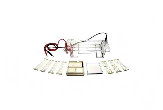
ال نظام الهلام الأفقي HE-50 من شركة لونغلايت تكنولوجي، يستخدم تصميما موحدا مفيدا لتحليل الرحلان الكهربائي للحمض النووي البلازميدي.
1. فصل عالي الجودة
•أقطاب بلاتين نقية بنسبة 99.95٪: عنصر تصميم حيوي لحل الأقطاب الملفوفة من الأزيوفورم المكسورة.
• تنسيق الجل المرن: يمكن للباحثين اختيار أبعاد الجل بأي من أحجام الصينية الأربعة المتاحة: 60×60 مم، 60×120 مم، 120×60 مم، و120×120 مم. الحجم الأكبر يوفر مسافة هجرة طويلة بما يكفي لرؤية الأشكال المتساوية المهاجرة بشكل قريب بدقة أعلى.
2. التصميم الموجه نحو المستخدم
• صينية تحميل موجهة بخلفية سوداء: المسطرة الفلورية ذات الخلفية الداكنة تتيح محاذاة رأس الماصة بعناية مع الآبار عند تحميل عدة عينات للمقارنة المباشرة.
• خزان البولي كربونات عالي الشفافية: تصميم عالي الرؤية مع خزان مدمج يظهر هجرة مقدمة الصبغة وعملية الرحلان الكهربائي دون الحاجة لفتح الخزان وقطع النظام.
3. السلامة والكفاءة
• غطاء الإغلاق التلقائي: يقوم مفتاح الأمان بقطع الطاقة عند فتح الغطاء لإزالة خطر الصدمة الكهربائية أثناء تحميل المستخدمين للعينات أو إزالة الجيلاتين.
• استهلاك منخفض للعازلة: تحتوي غرفة الهجرة السفلية على تصميم جسر يستخدم حاجز أقل مع توفير بيئة أيونية كافية لإجراء الرحلان الكهربائي للحمض النووي بشكل متسق وموثوق.
خطوات تحليل أشكال البلازميدات
للحصول على الفصل الأمثل للحمض النووي البلازميدي باستخدام نظام HE-50:
- اصنع جل الأغاروز: اختر أيا من أحجام الصينية الأربعة لصب جل أغاروز بنسبة 0.8-1.0٪. حلل البلازميدات التي تتراوح حجمها بين 3 - 10 كيلو باط باستخدام جل أغاروز مخزن ل 1× TAE.
- تحضير العينة: ادمج عينات الحمض النووي البلازميدي مع صبغة التحميل. استخدم 200 - 500 نانوغرام من حمض نووي البلازميد. نسب عينات الصبغة: صبغة التحميل يمكن أن تكون 1:6، 1:3، أو 1:1.5. باستخدام ماصة، قم بتحميل عينات صبغة البلازميد في آبار الأغاروز.
- الرحلان الكهربائي: يجب تشغيل جل بأبعاد 120 × 120 مم عند 100 فولت. بالنسبة للجل 100 × 100 مم، استخدم 80 فولت. يسمح للبروموفينول الأزرق بالعمل لمدة 45-60 دقيقة. يتوقف الرحلان الكهربائي عندما يصل الصبغة إلى ثلثي الطريق إلى القاع.
- تصور وتوثيق: الغلاف الشفاف المكشوف يسمح بتصوير الجل بالأشعة فوق البنفسجية وتصويره بينما الغلاف لا يزال في مكانه. عندما يكون هناك مسطرة جزيئية، يلاحظ الفلورة الإيجابية.
- قياس توزيع الأزوفورم: تقوم أنظمة التوثيق الهلامية الآلية بقياس شدة النطاق. يتم تحديد نسبة الحمض النووي الملفوف بالنحو التالي: [شدة SC] / (SC OC شدات خطية) × 100.
تكامل سير العمل دون عوائق
تم تصميم نظام HE-50 للدمج دون عوائق في سير عمل المختبرات الحالية:
•دمج مزود الطاقة: مقابس الموز المطلوبة بقطر 4 مم متوافقة مع معظم مصادر الطاقة الرحلان الكهربائي (نوصي بالجهد الثابت).
• دمج نظام التصوير: الغطاء الشفاف للأشعة فوق البنفسجية متوافق مع أنظمة تصوير التوثيق الهلامية القياسية، مما يلغي الحاجة لنقل الجل من الخزانات إلى أجهزة التصوير.
• اعتماد البروتوكول: النظام متوافق مع جميع تركيزات هلام الأغاروز القياسية، ومخازن الرحلان الكهربائي (TAE, TBE, SB)، مما يتيح اعتماد بروتوكولات تحليل البلازميدات المنشورة سابقا.
الخاتمة
الرحلان الكهربائي للحمض النووي هو الطريقة الأكثر سهولة وشعبية لتقييم جودة حمض البلازميد. يمكن للباحثين الاستفادة من معرفتهم بهجرة الأشكال الفائقة الملفوفة والمتشابكة والخطية لتحسين الفصل وتفسير النتائج. يوفر نظام HE-50 للجل الأفقي القدرات — أقطاب البلاتين النقي، أحجام الجل القابلة للاختيار، أدلة التحميل، وضوابط — الضرورية لتحليل البلازميدات الروتيني. كما ورد في مجلتي Nature Communications (2025) وNIH (2025)، فإن الرحلان الكهربائي بجل الأغاروز هو أحد الطرق الأساسية لتحليل التفاعلات بين الحمض النووي والبروتينات وكذلك طوبولوجيا أو بنية الحمض النووي.
نظام HE-50 الأفقي للهلام لديه القدرة على أن يكون متكاملا بالكامل وقابلا للتعديل مع أنظمة التصوير؛ وبهذه الطريقة، توفر للعملاء خيارات أكثر لأنظمة تصوير الحمض النووي البلازميدية بأسعار معقولة. لمعرفة المزيد، يرجى إرسال طلب عرض سعر إلى شركة لونغلايت تكنولوجي.
الأسئلة المتداولة
س1. هل يمكن للرحلان الكهربائي الهلامي الأفقي تحديد الحمض النووي الفائق الملفوف والمضغوط في البلازميد؟
يمكن لنظام HE-50 تحديد بلازميدات الحمض النووي الملفوفة والمحفوفة والخطية طالما تم تحقيق توحيد المجال الكهربائي، وتستخدم هلام الأغاروز بنسبة 0.8-1.2٪.
س2. ما هي معدلات الهجرة المختلفة لأشكال البلازميدات المختلفة أثناء الرحلان الكهربائي؟
نظرا لاختلاف أشكال البلازميدات من الناحية الهيكلية، فإنها تظهر معدلات هجرة متفاوتة عند تعرضها للرحلان الكهربائي. على سبيل المثال، تنتقل أشكال البلازميدات الخطية أسرع من الأشكال الدائرية. الحمض النووي في الشكل الخطي له معدل هجرة متوسط، بينما الحمض النووي في الشكل الدائري المقشر أو المفتوح لديه أبطأ معدل هجرة.
س3. ما هي نسبة الحمض النووي الفائق الملفوف التي تعتبر عالية الجودة للنقل؟
يعتبر الشكل الفائق الملفوف من حمض البلازميد النووي الذي يزيد حجمه عن 80٪ عادة تحضيرا عالي الجودة للبلازميد للنقل والعلاج الجيني.
س4. هل يمكنني استخدام مخزن TAE بدلا من TBE لفصل نظائر البلازميد؟
نعم، في معظم الحالات يمكن استخدام مخزن TAE لتحليل البلازميدات. ومع ذلك، فإن مخزن TBE هو الأمثل لفصل أشكال البلازميد عندما تكون الفروقات طفيفة وفي فترات ممتدة.
س5. لماذا تظهر عينات البلازميد لدي عدة شرائط على الجل؟
عادة ما تلاحظ وجود نطاقات متعددة للأشكال الملفوفة الفائقة، والمحفوزات، والخطية من البلازميد. قد تكون الأشرطة الأخرى نتيجة لسوء الهضم أو تدهور تلوث الحمض النووي والحمض النووي الريبي.
