

الوظائف ذات الصلة
التصوير المقطعي بالإلكترون الكريو-إلكترون (Cryo-ET): هل عينتك جاهزة لدراسة الأحياء الهيكلية في الموقع؟
2026-08-17أكثر تطبيقات التصوير المقطعي بالتبريد الإلكتروني ليست فقط "كيف يبدو هذا البروتين؟" بل أيضا: "كيف يبدو هذا الهيكل حيث يعمل فعليا؟"
هذا التمييز مهم. يمكن للمجمع المنقى أيضا أن يوفر مستوى مثاليا من التفاصيل الهيكلية مع إزالة الأغشية أو المجمعات المجاورة أو بنية الخلايا أو البنية الفيروسية المهمة للسؤال البيولوجي. يتعامل Cryo-ET مع هذا التحدي بطريقة مختلفة، من خلال أخذ العديد من الإسقاطات لعينة زجاجية لإعادة بناء حجم ثلاثي الأبعاد.
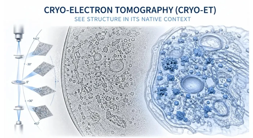
حاليا، تقدم لونغلايت تقنية Cryo-ET كنهج بنيوي ميداني للتجميعات البيولوجية، والمجمعات المرتبطة بالغشاء، والأنظمة الفيروسية كجزء من مشروعها خدمة المجهر الإلكتروني البارد.
السؤال العملي، مع ذلك، لجمع البيانات هو:
هل السؤال البيولوجي فعلا يتطلب إجراء تصوير طويل؟
خذ السؤال البيولوجي قبل المجهر
أحيانا، يشبه العلماء تقنية التبريد البارد بتحليل الجسيم الواحد، مما يشير إلى أن أحدهما كان يجب أن يكون متفوقا على الآخر.
عادة لا تكون مقارنة مباشرة بتفاح.
قد يكون التجمد المغناطيسي الجسيم الواحد مسارا أكثر مباشرة إلى بنية متوسطة عالية الدقة إذا كان مركب جزيء كبير منقى متجانس نسبيا يستطيع الإجابة على السؤال البيولوجي. ومع ذلك، يصبح Cryo-ET جذابا جدا إذا كان الموقع/الاتجاه، الجوار الجزيئي، بيئة الغشاء أو تنظيم الخلية للهدف مهما.
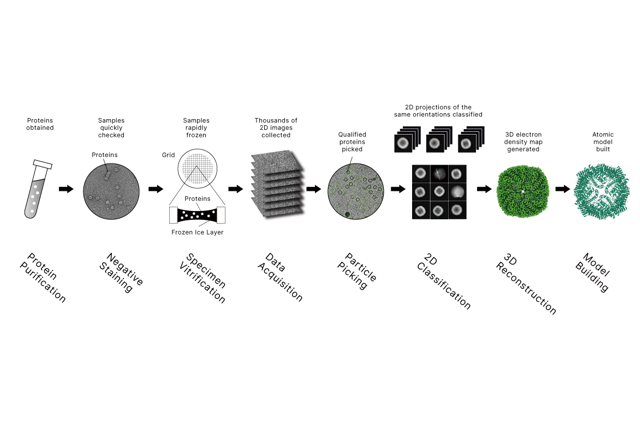
Longlight's دليل لتقنية Cryo-EM ذات الدقة القريبة من الذرة يقدم بعض الخلفية لمسار الجسيم الواحد؛ يطرح Cryo-ET سؤالا آخر: ماذا يمكن رؤيته عندما يتم الحفاظ على السياق البيولوجي؟
| هدف البحث | مسار بداية أكثر ملاءمة | سؤال رئيسي قبل التحصيل |
| البنية الجزيئية النقية والمتجانسة | التجميد الكهربائي الجسيم الواحد | هل يمكن للتطهير الحفاظ على الحالة المعنية؟ |
| تنظيم الخلايا الأصلية | كريو-إي تي | هل السياق المكاني ضروري؟ |
| المركب المرتبط بالغشاء في الموقع | كريو-إي تي | هل يمكن الوصول إلى المنطقة المستهدفة وتحديد موقعها؟ |
| المجمعات المتكررة داخل التصوير المقطعي | متوسط التصوير الفرعي للكريو-إي تي | هل تتوفر نسخ مماثلة كافية؟ |
| المنطقة الخلوية الداخلية السميكة | كريو-فيب كريو-إي تي | هل يمكن لللاميلا المناسبة أن تحتوي الهدف؟ |
الحد الفيزيائي الأول—إمكانية الوصول للإلكترونات
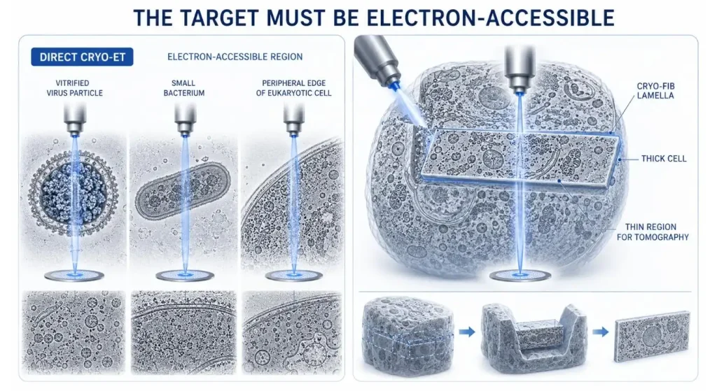
إذا كانت العينة مثيرة للاهتمام بيولوجيا، فهذا لا يعني بالضرورة أنها عينة يمكن الوصول إليها عبر الإلكترونات.
قد يكون من الممكن إجراء تصوير مقطعي مباشر بالتبريد للفيروسات أو العضيات المعزولة الأخرى، والخلايا الصغيرة، والمناطق المحيطية الرقيقة من الخلايا. تشمل بعض سير العمل في الموقع تخفيف العينات، وهو أمر ضروري لتصوير النقل المفيد للخلايا اليوسينوية الكبيرة التي تكون سميكة جدا بحيث لا يمكن تصويرها.
إحدى الطرق المعروفة لصنع صفائح رقيقة للخلايا هي الطحن بأشعة الأيونات المركزة بالتجميد (FIB). تم استخدام الخلايا حقيقية النواة المطحونة بالبرد الصناعي للشجرات في الدراسات الميدانية لجعل البنى الداخلية متاحة للتصوير المقطعي والتحليل البنيوي اللاحق.
يجب ألا يبدأ تخطيط العينات بجهد المجهر! يجب أن يشمل 'من أين' و'كيف'، سيصل شعاع الإلكترون إلى الهدف.
سلسلة تيلت ليست كومة من الصور، بل هي جرعة ميزانية.
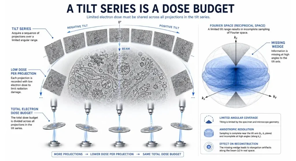
كريو-إي تي لا يحصل على معلومات عن البعد الثالث من تعريض واحد.
تميل هذه العينة عدة مرات ويتم التقاط سلسلة من الإسقاطات ثنائية الأبعاد. ويرجع ذلك إلى حساسية الإشعاع لدى العينات البيولوجية المجمدة، ويجب إعطاء الإسقاطات ميزانية محدودة لجرعة الإلكترون. كما يزداد السماكة الفعالة التي تمر بها الإلكترونات عند زوايا ميل أكبر، مما يعني أن إشارة أقل قد تكون قابلة للاستخدام.
في التصوير المقطعي التقليدي، لا يمكن الحصول على زاوية ±90° كاملة بسبب الهندسة الميكانيكية وسمك العينة. المعلومات غير المأخوذة من عينات تخلق الوتد المفقود المعروف في فضاء فورييه.
لكن المزيد من الإسقاطات لا يعني بالضرورة الأفضل. يجب أخذ التفاعل بين مدى الإمالة، توزيع الجرعة، جودة المحاذاة، استقرار العينة، ورؤية الهدف في الاعتبار.
حدد إذا كنت بحاجة إلى تصوير مقطعي، وهو هيكل متكرر.
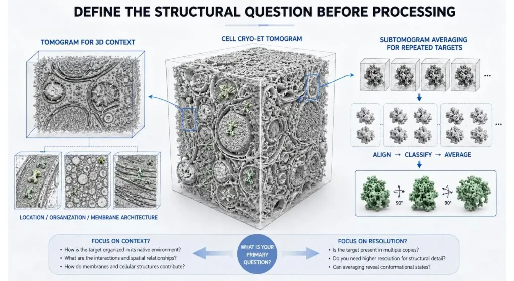
ليس كل مشاريع Cryo-ET يجب أن تنتهي بنموذج جزيئي شبه ذري!
غالبا ما يوفر التصوير المقطعي وحده إجابة على السؤال: أين يقع الهيكل؟ ماذا يحدث للغشاء عندما يتشوه؟ ما هو تنظيم مكونات الفيروس؟ أي المجمعات متجاورة لبعضها البعض؟
سيتم عرض هدف مختلف عندما تكون هناك نسخ متعددة من نفس المركب الجزيئي الكبير. يمكن استخراج الأهداف المتكررة كصور تحت التوموغرام، ومحاذاة وتصنيفها وقياسها لتعزيز الإشارة الهيكلية.
في هذا المقال، يقدم لونغلايت شرحا مفصلا لهذا المسار. القضية الرئيسية لمشروع جديد يتم التأكيد عليها من البداية: هل هذه بنية ثلاثية الأبعاد فريدة أم هناك نسخ مماثلة كافية للمتوسط؟
العنق الخفي يمكن أن يتحول إلى تحديد موقع الأهداف.
اختيار أفضل منطقة خلوية قد يكون مهما بقدر أهمية الحصول على سلسلة إمالة تقنية جيدة.
افترض أن الهدف هو جزء صغير فقط من خلية كبيرة. حتى لو كان لديك صورة مقطعية جميلة من المنطقة الخطأ، فهي تظل مجموعة بيانات خاطئة.
لكي يتم جمعها، يجب أن تأخذ في الاعتبار وفرة الهدف، وتحديد الموقع التقريبي للخلايا، وشكله المعروف، والمعلومات المتاحة حول الوسم، وما إذا كانت هناك حاجة لاستراتيجية تحديد مكان أخرى.
يمكن لهذه الخريطة البيولوجية أن تعزز بشكل كبير منطق خطة الاستحواذ للأهداف المرتبطة بالغشاء، ومواقع تجميع الفيروس، والعضيات، أو المجمعات المقيدة مكانيا.
التخطيط قبل الجلسة لمسار البيانات:
عندما يتم الحصول على سلسلة ميلت، لا يكتمل مشروع Cryo-ET.
يجب بعد ذلك محاذاة الصور، وإعادة بناؤها إلى مخططات مقطعية، وفحصها، والبحث عن أهداف، وتقسيمها، وإذا كان ذلك مناسبا، نقلها إلى استخراج الجسيمات والتصنيف ومتوسط التصوير الفرعي.
لهذا السبب يجب تحديد المنتج المطلوب مسبقا للجمع.
في خدمة Cryo-EM الحالية من لونغلايت، نركز على تسليم خرائط الكثافة النهائية، وكذلك على البيانات الخام وملفات المعالجة الوسيطة. وهذا مفيد عندما يرغب الباحث في التحقق من قرارات المعالجة، أو إجراء تحليلات لاحقة داخل النظام، أو العودة إلى مجموعة البيانات في وقت لاحق.
| معلومات المشروع | ماذا يجب أن تجهز |
| نوع العينة | الخلايا، الفيروسات، العضيات، الأغشية، التجمعات المنقاء |
| موقع الهدف | السيتوبلازم، الغشاء، العضية، المنطقة الفيروسية، موقع آخر |
| وفرة الأهداف | التردد المتوقع أو توزيع النسخ التقريبي |
| الهدف الهيكلي | السياق ثلاثي الأبعاد، الشكل، STA، إعادة بناء الجزيئات |
| الأدلة الحالية | بيانات التألق، الكيمياء الحيوية، الEM، أو البيانات الهيكلية |
| المخرجات المطلوبة | البيانات الخام، التصوير الطوتوغرافي، الخرائط المعالجة، النماذج، التقارير |
منصة المطابقة إلى مرحلة المشروع
لا ينبغي استخدام جودة الأداة لمعالجة سؤال عينة غير محدد.
يمكن أن يشير الفحص الأولي إلى ما إذا كان يجب تحسين تحضير العينة، توزيع الجزيئات، جودة الجليد، و/أو رؤية الهدف قبل استثمار وقت جمع مكلف.
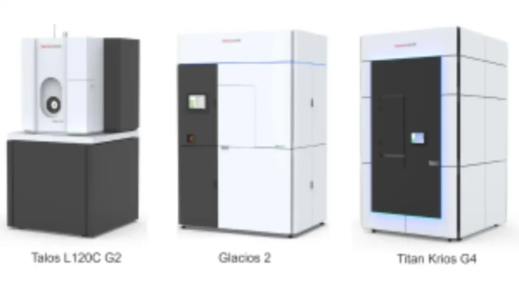
أعلنت شركة لونغلايت اليوم عن منصة تجميد مغناطيسي على مراحل تتكون من Talos L120C G2 للفحص، وGlacios 2 بجهد 200 كيلوفولت للتصوير المقطعي بالتجميد الروتيني/التصوير المقطعي، وTitan Krios G4 بجهد 300 كيلو فولت لشراء الأجهزة عالية المستوى المطلوبة. كما يرتبط بسير عمل البروتين في المراحل العليا من خلال أوسع تطبيق البروتين محفظة تربط التحليل الهيكلي بمحفظتها.
قاعدة البحث العامة هي:
العينة جاهزة، انتقل إلى الآلة التالية، وليس لأنها متوفرة.
هذا هو الفحص النهائي لمشروع Cryo-ET.
فيما يتعلق بتخصيص عينة للتصوير المقطعي الإلكتروني التبريدي، هناك خمسة أسئلة يجب أخذها في الاعتبار:
هل هناك حاجة إلى سياق مكاني أصلي؟ هل منطقة الاهتمام في الجزيء متاحة للإلكترون؟ هل هناك رؤية موثوقة للهدف؟ أوه، تصوير مقطعي واحد أم جزيئات متكررة؟ هل تعرف نوع منتجات البيانات التي ستكون مطلوبة بعد الاستحواذ؟
بمجرد تحديد تلك الإجابات، يصبح اختيار المعدات واستراتيجية المعالجة أسهل بكثير.
تكمن قوة التجميد المبكر في أنه يحافظ على المعلومات التي تفقد أثناء التنقية. ومع ذلك، سيكون هذا مفيدا فقط إذا تم تصميم تحضير العينة، واستراتيجية التصوير، والسؤال البيولوجي معا كمشروع واحد.
الباحثون يمكنهم استشارة لونغلايت في مشروع هيكلي ميداني، بعد أن أصف لهم نوع العينة التي سيستخدمونها، والموقع المستهدف، ونوع الهيكل الذي يسعون لتحقيقه، والتحليل الذي سيستخدمونه في المراحل النهائية.
الأسئلة المتداولة
س1. تصوير مقطعي بالتبريد الإلكتروني (Cryo-ET)؟
يتضمن Cryo-ET التقاط عدة صور لعينة بيولوجية في الفيترين من زوايا مختلفة ثم إعادة بنائها إلى تصوير مقطعي ثلاثي الأبعاد. إحدى الفوائد الرئيسية هي أنه يحافظ على المعلومات البنيوية في بيئة بيولوجية شبه أصلية.
س2. ما هي الظروف المناسبة لاستخدام Cryo-ET فوق جسيم واحد من التجميد الكهرومغناطيسي؟
فكر في التجميد المغناطيسي عندما يكون موقع الخلية، أو تنظيم الغشاء، أو الجيران الجزيئيين، أو البنية الأصلية عاملا رئيسيا في سؤال البحث. عندما يكون التركيز على الجسيم المنقي/البنية الجزيئية المتوسطة المتوسطة، يكون SPA غالبا أكثر مباشرة.
س3. هل يجب استخدام طحن التجميد والرجفان الصناعي لجميع عينات Cryo-ET؟
لا. من الممكن تصوير عينات مخففة طبيعيا مباشرة. في المناطق الأكثر سمكا من الخلية، قد يكون من الضروري تخفيف التخفيف لإنشاء جزء شفاف من الإلكترونات من الخلية لإجراء التصوير المقطعي.
س4. ما الذي ينقص في Cryo-ET؟
يفتقر إلى معلومات ثلاثية الأبعاد من النطاق الزاوي المحدود الذي يمكن الحصول عليه أثناء سلسلة الإمالة. قد يسبب عدم التوافق، أي تشوهات إعادة البناء، والتي يجب أخذها في الاعتبار أثناء التحليل.
س5. هل يجب إجراء متوسط التصوير الفرعي لكل مشروع يتم تنفيذه في Cryo-ET؟
لا. يمكن أن يكفي مخطط مقطعي واحد للإجابة على أسئلة تتعلق بالشكل أو التنظيم المكاني. تكمن قيمة المتوسط الفرعي في إمكانية محاذاة ومتوسط عدة نسخ متشابهة من الهدف عندما تكون متاحة.
