

الوظائف ذات الصلة
اكتشاف الحمض النووي الريبي منخفض الوفرة: كيف تلتقط منصة تصوير هلام RNA عالية الحساسية النطاقات الضعيفة
2026-06-23تحدي تصور جزيئات الحمض النووي الريبي النادرة
في أبحاث علم الأحياء الجزيئي، يطرح تحليل الحمض النووي الريبي تحديات فريدة. الحمض النووي الريبي أكثر عدم استقرار من الحمض النووي النووي، لذا فهو أصعب في التعامل، حيث يتحلل بسهولة وغالبا ما يكون موجودا بكميات أقل. وهذا ينطبق على العديد من الفئات المهمة من الحمض النووي الريبي، مثل ال miRNAs، أو الحمض النووي الريبي غير المشفر، أو نسخ RNA ذات التعبير المنخفض. نظرا لصعوبة التعامل مع هذه الجزيئات، فمن الضروري أن تكون لديك القدرة على رؤية هذه الجزيئات الكبيرة.
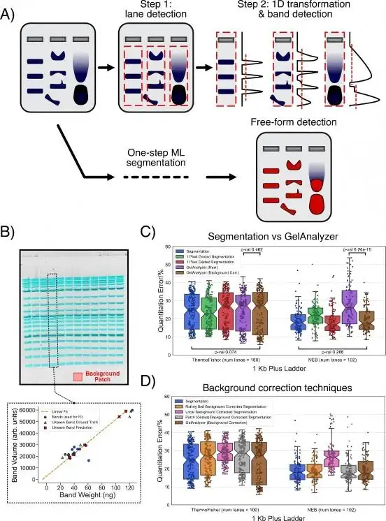
[GelGenie: إطار عمل مدعوم بالذكاء الاصطناعي لتحليل صور الرحلان الكهربائي بالجل]
وتؤكد هذه الصعوبة بعض التطورات الحديثة الناتجة عن الظاهرة التي وصفت سابقا. في عام 2025، قدم باحثون من بينهم م. أكويلينا جهاز GelGenie. GelGenie هو محلل صور يدوي بالجل. يتضمن مع GelGenie محلول تصوير مصمم لإزالة الأعمال اليدوية من عملية التصوير. في عام 2026، وصف ي. تشينغ والمؤلفون طريقة شديدة الحساسية للكشف عن الحمض النووي الريبي الورمي الدائر منخفض الوفرة (ctRNA) من عينة دم في منشور لمجلة Nature Communications. أفادت دراستهم بمستوى حساسية مطلوب يبلغ 0.059 جزء في المليون.
لماذا يعتبر اكتشاف نطاقات RNA منخفضة التعبير معقدا؟
تساهم عدة عوامل في صعوبة تصور نطاقات RNA. إليك كيف سأشرح ملاحظاتك.
• الكمية غير كافية: مستويات النسخ منخفضة وبالتالي مستويات الفلورسنت منخفضة أيضا وقد لا يمكن اكتشافها بواسطة نظام التصوير.
• خلفية عالية: عندما تكون إشارة الفلورة الإلكترونية ضعيفة، قد تخفي إشارة الحمض النووي الريبي بواسطة التألق الخلفي والتلوين غير النوعي.
• تجزئة الحمض النووي الريبي: يعتبر إنزيم RNase من أكثر الإنزيمات غير المحددة والملوثة من خطوات تحضير العينة، ويمكن أن يحلل الحمض النووي الريبي ويسبب إشارة RNA مجزأة أو مشوشة عبر عدة نطاقات.
• الصبغات: تتطلب صبغات الحمض النووي الريبي مطابقة لطول موجة الإثارة الخاص بالصبغة المستخدمة للحصول على إشارة قوية.
• منصة تصوير هلام RNA عالية الأداء مع أجهزة خاصة وتصميم مدروس هي الحلول لهذه المشاكل.
التقنيات الرئيسية لاكتشاف الحمض النووي الريبي
1. كاميرا CMOS عالية الحساسية
أنظمة الكشف هي أساس منصات تصوير الحمض النووي الريبي الحديثة للهلام. كاميرات CMOS علمية الجودة، مع:
• 6.3 ميجابكسل (MP) لتفاصيل النطاقات الدقيقة
• 66 ديسيبل من الإشارة إلى الضوضاء لاكتشاف الإشارة الحقيقية وسط الضوضاء
• انخفاض مستويات التعريض للأشرطة الخلفية المعرضة بشكل مفرط
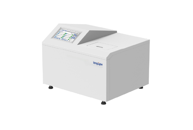
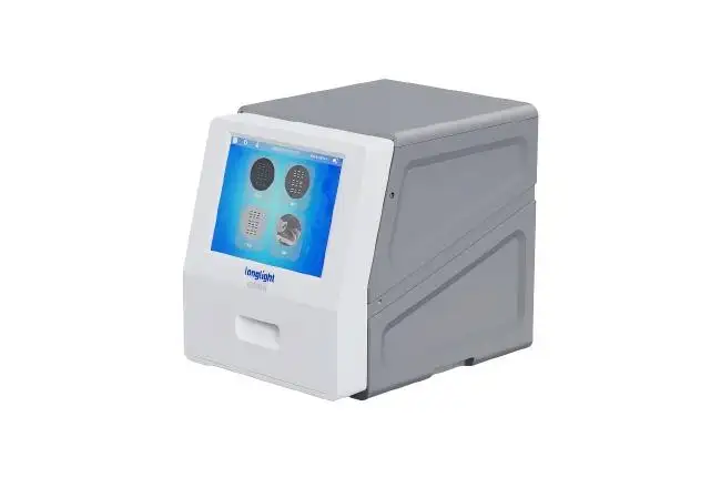
يستخدم نظام التصوير الهلام GI-200 من شركة Longlight Technology كاميرا CMOS عالية الحساسية بدقة 6.3 ميجابكسل ويوفر نطاقات واضحة حتى في ظروف الإضاءة المنخفضة.
2. إثارة LED متعددة المصادر
هناك مقايضة بين صبغات RNA المختلفة وأقصى إثارتها. يجب أن يقدم النظام متعدد الاستخدامات والمتكامل:
• الأشعة فوق البنفسجية (302 نانومتر): إيثيديوم بروميد وجيل ريد
• ترانس-بلو (470 نانومتر): SYBR Green II وSYBR Safe أكثر أمانا للمستخدم وللعينة
• التحول إلى الأبيض: صبغات البروتين اللونية وعد المستعمرات
يضمن هذا النظام الإثارة المثلى لأي صبغة RNA.
اختراقات حديثة في اكتشاف الحمض النووي الريبي منخفض الوفرة
شهدت الريانا تطورات سريعة في حساسية اكتشاف الحمض النووي الريبي.
1. طريقة ستالارد (2025)
طريقة ستالارد (التضخيم الانتقائي للهدف لاكتشاف الحمض النووي الريبي منخفض الوفرة) هي طريقة RT-PCR ذات خطوتين لتضخيم الحمض النووي الريبي المستهدف المفضل بين الحمض النووي الريبي متعدد الأدينيل الذي يشترك في تسلسل نهاية 5 أقدام. تمكنت طريقة STALARD من اكتشاف نسخة VIN3 منخفضة الوفرة ونسخة COOLAIR الأقل حتى في Arabidopsis thaliana وحل المشكلات من الدراسات السابقة.
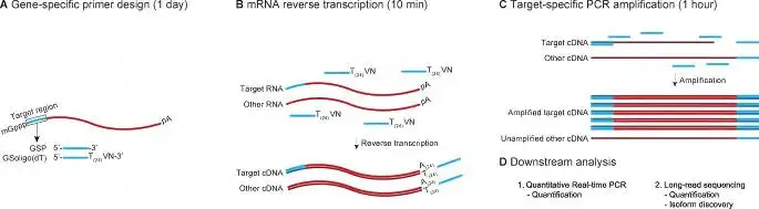
[ستالارد: تضخيم الهدف الانتقائي للكشف عن RNA منخفض الوفرة | طرق النبات | رابط سبرينغر نيتشر]
2. منصة SE-SPTM-PCR (2026)
في دراسات حديثة، طور منشور في مجلة Nature Communications (2026) من تأليف M. Aquilina وآخرين، منصة SE-SPTM-PCR، التي تجمع بين إثراء miRNA الانتقائي مع المسبار الانتقائي النهائي RT-qPCR. حقق هذا النظام حساسية أعلى بمقدار 100 مرة من RT-qPCR التقليدي ذو الحلقة الجذعية. في الدراسات السريرية، حسن التحليل الكيميائي للكشف عن سرطان القولون والمستقيم باستخدام has-miR-92a-3p من 0.72 إلى 0.85 عبر 96 مريضا.
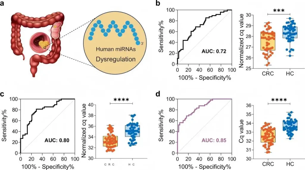
[استراتيجية تخصيب انتقائي واستراتيجية موجهة بجانب المسبار المحدد ل
الكشف الحساس جدا للميكروRNA | نيتشر كوميونيكيشنز]
تؤكد هذه التطورات نقطة حاسمة: يجب أن تتوافق حساسية طريقة الكشف مع حساسية نظام التصوير. حتى أفضل استراتيجية تضخيم تتطلب تصورا موثوقا.
كيف يعزز GI-200 تصور الحمض النووي الريبي منخفض الوفرة
تقنية Longlight'نظام تصوير الجل s GI-200 تتضمن ميزات تصميم مفيدة بشكل خاص لتطبيقات الحمض النووي الريبي.
1. معالجة الصور الذكية
يدعم البرنامج الذي تم تطويره ذاتيا ما يلي:
• التعريض التلقائي: يحسن زمن التقاط الإشارة بناء على شدة الإشارة
• التركيز التلقائي: يلغي التفاوت بين المستخدمين
• تحسين الصورة: يحسن التباين دون تشويه البيانات الكمية
كل ما يفعله المستخدمون هو النقر على زر الصورة، مما يقلل من وقت معالجة البيانات ويحسن الكفاءة.
2. التكامل الشامل
يتميز GI-200 بشاشة لمس مقاس 12.1 بوصة مع معالجة صور مدمجة، مما ألغى الحاجة إلى حاسوب خارجي. هذا يوفر المساحة والوقت أثناء المعالجة. وهو مفيد بشكل خاص للعمل مع الحمض النووي الريبي لأن خطر التلوث أعلى.
3. السلامة لاستئصال الجل
السلامة أمر بالغ الأهمية عند إزالة نطاقات RNA لاستخدامها في RT-qPCR، والتسلسل، والاستنساخ، وغيرها:
• يدعم قطع الجل الخارجي لاستخراج الشريط بدقة أكبر
• يغطي لوحة قطع مقاومة للأشعة فوق البنفسجية لحماية المستخدمين من التعرض للأشعة فوق البنفسجية الخطرة
• متوافقة مع وضع الضوء الأزرق لرؤية أكثر أمانا وعدم ضرر للعينات التي ستخضع لاختبارات وظيفية
نصائح عملية لتحسين تصوير جل الحمض النووي الريبي
لضمان أقصى إمكانات النجاح عند استخدام منصة تصوير الجل RNA، ضع في اعتبارك ما يلي:
| الاعتبار | التوصية |
| سلامة الحمض النووي الريبي | دائما استخدم جل تغيير الطلاء للتحقق من نسبة rRNA 28S/18S قبل التطبيقات النهائية |
| اختيار البقع | استخدم SYBR Green II أو SYBR Safe مع إثارة الضوء الأزرق بدلا من EtBr لأن الأخير له حساسية أقل |
| زمن التعريض | بالنسبة للأشرطة الضعيفة، ابدأ بتعريض أطول؛ تحسين باستخدام ميزة التعريض التلقائي |
| تقليل الخلفية | إزالة الصبغة واستخدام أنظمة تصوير عالية الإشارة إلى الضوضاء |
| الوثائق | استخدم أنظمة تحتوي على مسار تدقيق للامتثال لمعايير GLP/GMP |
خط أنابيب تصوير الحمض النووي الريبي منخفض الوفرة
تشير أبحاث جديدة إلى ما يلي:
• تحليل أنظمة الذكاء الاصطناعي: يمكن ل GelGenie إجراء اكتشاف أشرطة الجل تلقائيا في مجموعة واسعة من الإعدادات التجريبية
• التصوير متعدد الإرسال: تسمح أنظمة SATA-FISH بالكشف الحساس عن تسلسلات RNA القصيرة في أنسجة FFPE
• دقة جزيء واحد: تسمح مجسات تقرير FRET المعتمدة على QD بالكشف عن mRNA داخل الخلية عند 35 fM
مع تقدم هذه الأنظمة، سيكون هناك زيادة مماثلة في الطلب على منصات تصوير هلام RNA عالية الحساسية.
استنتاج
لا يزال اكتشاف الحمض النووي الريبي منخفض الوفرة يواجه العديد من الصعوبات في علوم الحياة. يمكن للتطورات التي تجمع بين الكيمياء الحساسة للكشف والتضخيم القوي والتصوير المتقدم الآن من الممكن اكتشاف وقياس أندر النسخ.
يعد نظام التصوير الهلامي GI-200 من شركة لونغلايت تكنولوجيز من أوائل أنظمة التصوير التي صممت مع مراعاة احتياجات أبحاث الحمض النووي الريبي الحالية. من خلال دمج كاميرا CMOS عالية الحساسية بدقة 6.3 ميجابكسل، ومصادر إثارة متعددة، وأتمتة تصوير متقدمة، يوفر GI-200 كل ما يحتاجه المختبر لتوسيع إمكانيات أبحاث الحمض النووي الريبي.
هل تود رؤية GI-200 أثناء العمل؟
[طلب اقتباس مجاني] | تجربة الفوائد | حمل المواصفات
الأسئلة الشائعة
س1. هل يمكنني استخدام نفس نظام تصوير جل الحمض النووي الريبي لجل الحمض النووي أيضا؟
بالطبع. يمكن لأنظمة التصوير الحديثة التبديل بين الحمض النووي الريبي وهلام الحمض النووي عن طريق تغيير مصدر الإثارة وصبغة التصوير.
س2. ما هي أقل كمية من الحمض النووي الريبي يمكن اكتشافها؟
مع كاميرا CMOS بدقة 6.3 ميجابكسل وإشارة إشارة ضوضاء بسرعة 66 ديسيبل، يمكن لأنظمة مثل GI-200 اكتشاف الحمض النووي الريبي على مستوى pg.
س3. هل الضوء الأزرق أفضل من الأشعة فوق البنفسجية في تصوير الحمض النووي الريبي؟
الضوء الأزرق هو مصدر إثارة أكثر أمانا وأقل ضررا للحمض النووي الريبي مقارنة بالأشعة فوق البنفسجية، خاصة للحمض النووي الريبي الذي يستخدم في التطبيقات اللاحقة.
س4. هل سأحتاج إلى جل تغيير الصبغة لتصوير الحمض النووي الريبي؟
نعم. تستخدم جل التغيير الفورمالديهايد أو اليوريا وتسمح بفصل دقيق حسب الحجم دون تركيب ثانوي RNA.
س5. كيف يعمل التعريض التلقائي للأشرطة الضعيفة من الحمض النووي الريبي؟
التعريض التلقائي يحسن زمن التعريض للإشارات التي يجب سحبها من الضوضاء، مع منع التعريض الزائد لمجال التصوير.
